|
|
|
||
| Home | About | Donate/Volunteer | Contact | Jobs| |
|
|
New dynamic imaging techniques provide a deeper look at the disease process of schizophrenia and Alzheimer's | ||||||||||
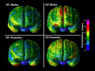
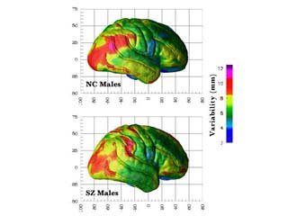
Using magnetic resonance imaging and a new analysis technique, researchers have created the first images showing the toll schizophrenia takes on the brain. (UCLA) By Robert Adler (Images added below, Source: Laboratory of Neuro Imaging, UCLA) The movie lasts just a few seconds, but in a few disturbing, color-coded frames it reveals what happens to the brains of Alzheimer's patients as they descend from worrisome memory lapses into dementia. Paul Thompson, the UCLA neuroscientist whose team created these first-ever sequences of a disease engulfing the living human brain, sees them as a significant step toward earlier diagnosis, more effective treatment, and -- eventually -- prevention or cure of brain-destroying diseases. Sid Gilman, director of the Michigan Alzheimer's Disease Research Center, concurred. ''The images are pretty dramatic,'' he said, ''and what they show is very important.'' With 10 million Alzheimer's cases expected in the United States by 2025, progress is vital. Thompson, a 31-year-old British emigrant, is unabashedly excited about his team's accomplishments: ''The tools from our group are opening really new windows on what's happening inside the brain.'' They give researchers a powerful way to test new medications and lets doctors diagnose Alzheimer's and other dementias earlier and more accurately. That should give more patients an early start on medications that can at least slow the ravages of these diseases. Like faces, no two brains are alike. As a result, it's extremely difficult to compare diseased and healthy brains or track changes over time. That's the problem Thompson and his team solved. By morphing ordinary MRI scans onto a standardized brain, they can pool scans from multiple patients without blurring the picture. They can then sequence those standardized images into revealing movies. And, crucially, they can quantify changes with great precision. ''We can code normal human variation,'' he said, ''and still be exquisitely sensitive to abnormal changes.'' As a result, Thompson's group has been able to study diseases such as Alzheimer's and schizophrenia as never before. ''With this kind of imaging, you can see a lava flow of destruction as more and more brain tissue is engulfed,'' Thompson said. ''You can see exactly which areas are losing tissue, when, and how fast.'' [While the healthy teens lost an average of 1 percent of gray matter per year, the schizophrenic patients lost up to 5 percent a year, with loss greatest among individuals with the most severe symptoms. - ed]
Thompson first applied these new tools to schizophrenia, a life-changing illness that strikes about one young person in a hundred worldwide. Researchers continue to debate schizophrenia's causes. It can devastate thinking, feeling and behavior without causing obvious brain damage. Some imaging studies showed thinning of the cortex, the brain's outer layer. Others saw a shrunken hippocampus -- a nubbin of tissue deep within the brain that is vital for storing memories. Until recently, these findings created more controversy than clarity. In 2001, Thompson's group produced the first time-lapse images revealing a wave of tissue loss rolling across the brains of schizophrenic children. They utilized high-resolution MRIs of more than 1,000 children scanned every two years since 1992 by Judith Rapoport and colleagues at the National Institute of Mental Health. Thompson's group detected the first flicker of the disease in a small part of the parietal cortex, above and behind the ears. Over five years, Thompson saw a ''pervasive, unrelenting wave of tissue loss that swept forward like a forest fire,'' eventually engulfing the sides and front of the brain. By 18, the teenagers had lost 25 percent of their gray matter in certain brain areas.
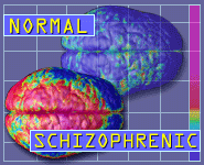
''Seeing that wave of tissue loss in schizophrenia was a huge surprise,'' Thompson said. The pattern matched the drumbeat of schizophrenia's active and passive symptoms -- hallucinations, delusions and bizarre thinking followed by flattened emotions, depression and withdrawal. The images are disturbing but valuable. They've pushed Thompson toward the theory that schizophrenia is a disrupted version of normal brain development. Teenagers' brains normally undergo extensive ''pruning'' in which 1 percent of the gray matter disappears every year, more in some areas. Because schizophrenia typically strikes during this process, Thompson sees it as ''an exaggeration or derailment'' of normal pruning -- like a gardener gone wild. His finding that schizophrenia takes up to seven years to engulf the brain highlights the need for early diagnosis and treatment. It also makes finding drugs that may salvage young people's brains even more vital. ''There is a window of opportunity to step in and oppose the disease,'' he said. Unlike schizophrenia, Alzheimer's usually strikes the elderly and often leads to death. Since pathologists have autopsied the brains of thousands of Alzheimer's patients, Thompson knew what his movies ought to reveal. As reported in the Journal of Neuroscience earlier this year, the tissue losses he found in living patients tracked the autopsy studies almost perfectly. The new images also made sense of the dreaded progression of Alzheimer's symptoms. Early memory and emotional problems matched damage deep within the brain. Erosion of higher mental functions mirrored a wave of damage that swept across the parietal and temporal lobes on both sides of the brain, inundating the language-processing left hemisphere first. By the time the disease engulfed patients' frontal lobes, their personalities and lives were shattered. Thompson thinks the findings offer a clue to what causes Alzheimer's. The wave of gray-matter loss he saw matches the spread of beta-amyloid plaques through the brain, supporting the theory that the build-up of that abnormal protein is a key to the disease. That makes treatments that target amyloid, including a controversial vaccine, look more promising. Although the new imaging technology is powerful, sophisticated and dramatic, it's just one step toward the prevention or cure of Alzheimer's, schizophrenia, and other brain-killing diseases. The work's first application may be in the early detection of these diseases, allowing more people to benefit from medications that slow the progress of schizophrenia and Alzheimer's. The new scans also will help researchers evaluate current and future drugs. Precision measurements of gray matter saved or lost gives drug researchers a clearer target to aim for. Dynamic brain imaging should also speed up the search for genes that predispose people to specific brain diseases. We know they're there. Siblings and children of schizophrenics have one chance in 10 of developing the disease -- 10 times the average risk. A half-dozen genes already have been linked to Alzheimer's, with more to come. It is much easier to match suspect genes to specific patterns of tissue loss than to shifting, hard-to-measure symptoms. Adam Boxer, a neurologist at the Memory and Aging Center of the University of California at San Francisco, ''I'm very excited by this technique,'' Boxer said. ''We know a lot about the molecular biology of Alzheimer's, but we don't understand on a millimeter-by-millimeter scale how it affects the living human brain.'' Thompson's revealing movies and the methodology behind them bring us closer to that goal. For a good sample of pictures comparing brains with schizophrenia and those without - see: Pictures of Schizophrenia: Neuro Imaging of the impact of the disease process of schizophrenia Related Story: Thompson's movies and additional images can be viewed at http://www.loni.ucla.edu/
|
Advertisement
|