|
|
||
|
|
|
|
|
Home
| About
| Donate/Volunteer
| Contact
| |
| Schizophrenia Information > Schizophrenia Pictures and Images of Brains | ||||||||||||
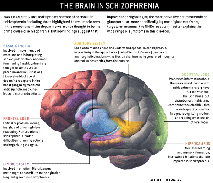
| Schizophrenia Pictures and Images of Brains |
||||||||||||
|
|
|
| |
|
|
This site does not provide medical or any other health care or fitness advice, diagnosis, or treatment. The site and its services, including the information above, are for informational purposes only and are not a substitute for professional medical or health advice, examination, diagnosis, or treatment. Always seek the advice of your physician or other qualified health professional before starting any new treatment, making any changes to existing treatment, or altering in any way your current exercise or diet regimen. Do not delay seeking or disregard medical advice based on information on this site. Medical information changes rapidly and while Schizophrenia.com makes efforts to update the content on the site, some information may be out of date. No health information on Schizophrenia.com, including information about herbal therapies and other dietary supplements, is regulated or evaluated by the Food and Drug Administration and therefore the information should not be used to diagnose, treat, cure, or prevent any disease without the supervision of a medical doctor.